SmartCT

3D visualization and measurement solution

Find similar products

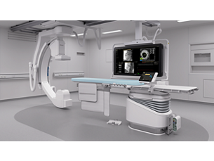
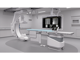
The SmartCT solution enriches our outstanding 3D interventional tools with clear guidance, designed to remove barriers to acquiring 3D images in the interventional lab. It simplifies 3D acquisition to empower all clinical users to easily perform 3D imaging, regardless of their experience[1]. Once acquired, 3D images are automatically displayed within seconds on the touch screen module in the corresponding rendering mode. On the same touch screen, the user can easily control and interact with advanced 3D visualizations and measurement tools.
Features
SmartCT empowers you to easily adopt 3D imaging in the lab

SmartCT empowers you to easily adopt 3D imaging in the lab

3D imaging can enhance diagnostic accuracy[2-4], improve patient outcomes[5-7] and increase procedural efficiency in the interventional lab[8]. Despite these advantages, it can still be considered difficult to perform by many users. To take the guesswork out of 3D acquisition, SmartCT provides step-by-step guidance and visual aids during acquisition to help any clinical user easily acquire 3D images.

SmartCT empowers you to easily adopt 3D imaging in the lab

SmartCT empowers you to easily adopt 3D imaging in the lab
3D imaging can enhance diagnostic accuracy[2-4], improve patient outcomes[5-7] and increase procedural efficiency in the interventional lab[8]. Despite these advantages, it can still be considered difficult to perform by many users. To take the guesswork out of 3D acquisition, SmartCT provides step-by-step guidance and visual aids during acquisition to help any clinical user easily acquire 3D images.

SmartCT empowers you to easily adopt 3D imaging in the lab

3D imaging can enhance diagnostic accuracy[2-4], improve patient outcomes[5-7] and increase procedural efficiency in the interventional lab[8]. Despite these advantages, it can still be considered difficult to perform by many users. To take the guesswork out of 3D acquisition, SmartCT provides step-by-step guidance and visual aids during acquisition to help any clinical user easily acquire 3D images.
Click here for more information
SmartCT empowers you to easily adopt 3D imaging in the lab

SmartCT empowers you to easily adopt 3D imaging in the lab

3D imaging can enhance diagnostic accuracy[2-4], improve patient outcomes[5-7] and increase procedural efficiency in the interventional lab[8]. Despite these advantages, it can still be considered difficult to perform by many users. To take the guesswork out of 3D acquisition, SmartCT provides step-by-step guidance and visual aids during acquisition to help any clinical user easily acquire 3D images.
Acquire and interact with 3D imaging at table side

Acquire and interact with 3D imaging at table side

With the touch screen, you can easily acquire 3D images and interact with SmartCT tools in a natural and effortless way. Once acquired, The SmartCT viewing application automatically opens your 3D image with the correct rendering and viewing tools on the touch screen module. All tools work with the tablet’s touch screen simplicity within the sterile area.

Acquire and interact with 3D imaging at table side

Acquire and interact with 3D imaging at table side
With the touch screen, you can easily acquire 3D images and interact with SmartCT tools in a natural and effortless way. Once acquired, The SmartCT viewing application automatically opens your 3D image with the correct rendering and viewing tools on the touch screen module. All tools work with the tablet’s touch screen simplicity within the sterile area.

Acquire and interact with 3D imaging at table side

With the touch screen, you can easily acquire 3D images and interact with SmartCT tools in a natural and effortless way. Once acquired, The SmartCT viewing application automatically opens your 3D image with the correct rendering and viewing tools on the touch screen module. All tools work with the tablet’s touch screen simplicity within the sterile area.
Click here for more information
Acquire and interact with 3D imaging at table side

Acquire and interact with 3D imaging at table side

With the touch screen, you can easily acquire 3D images and interact with SmartCT tools in a natural and effortless way. Once acquired, The SmartCT viewing application automatically opens your 3D image with the correct rendering and viewing tools on the touch screen module. All tools work with the tablet’s touch screen simplicity within the sterile area.
Control advanced 3D visualization and measurement tools at table
Control advanced 3D visualization and measurement tools at table side

Control advanced 3D visualization and measurement tools at table side

Using simple tablet gestures, you can carry out advanced measurements and visualizations on the touch screen at table side to study the type and extent of disease with great detail. SmartCT 3D images can help reveal information not apparent in 2D or DSA images. This additional information may change diagnosis, treatment planning or treatment delivery, supporting better patient outcome[8,9,10].

Control advanced 3D visualization and measurement tools at table side

Control advanced 3D visualization and measurement tools at table side
Using simple tablet gestures, you can carry out advanced measurements and visualizations on the touch screen at table side to study the type and extent of disease with great detail. SmartCT 3D images can help reveal information not apparent in 2D or DSA images. This additional information may change diagnosis, treatment planning or treatment delivery, supporting better patient outcome[8,9,10].

Control advanced 3D visualization and measurement tools at table side

Using simple tablet gestures, you can carry out advanced measurements and visualizations on the touch screen at table side to study the type and extent of disease with great detail. SmartCT 3D images can help reveal information not apparent in 2D or DSA images. This additional information may change diagnosis, treatment planning or treatment delivery, supporting better patient outcome[8,9,10].
Click here for more information
Control advanced 3D visualization and measurement tools at table
Control advanced 3D visualization and measurement tools at table side

Control advanced 3D visualization and measurement tools at table side

Using simple tablet gestures, you can carry out advanced measurements and visualizations on the touch screen at table side to study the type and extent of disease with great detail. SmartCT 3D images can help reveal information not apparent in 2D or DSA images. This additional information may change diagnosis, treatment planning or treatment delivery, supporting better patient outcome[8,9,10].
SmartCT Vessel Analysis
SmartCT Vessel Analysis with next generation vessel tracking supports treatment planning

SmartCT Vessel Analysis with next generation vessel tracking supports treatment planning

To quickly define a vessel path on a 3D volume, Select a start and end point of the segment of the vasculature of interest and the path between the 2 points is automatically detected and rendered in different views. SmartCT Vessel Analysis supports the selection of the optimal projection angle for vessel analysis and catheterization. It allows easy inspection of vessel and device positioning with straightened, curved and cross-section reformats.

SmartCT Vessel Analysis with next generation vessel tracking supports treatment planning

SmartCT Vessel Analysis with next generation vessel tracking supports treatment planning
To quickly define a vessel path on a 3D volume, Select a start and end point of the segment of the vasculature of interest and the path between the 2 points is automatically detected and rendered in different views. SmartCT Vessel Analysis supports the selection of the optimal projection angle for vessel analysis and catheterization. It allows easy inspection of vessel and device positioning with straightened, curved and cross-section reformats.

SmartCT Vessel Analysis with next generation vessel tracking supports treatment planning

To quickly define a vessel path on a 3D volume, Select a start and end point of the segment of the vasculature of interest and the path between the 2 points is automatically detected and rendered in different views. SmartCT Vessel Analysis supports the selection of the optimal projection angle for vessel analysis and catheterization. It allows easy inspection of vessel and device positioning with straightened, curved and cross-section reformats.
Click here for more information
SmartCT Vessel Analysis
SmartCT Vessel Analysis with next generation vessel tracking supports treatment planning

SmartCT Vessel Analysis with next generation vessel tracking supports treatment planning

To quickly define a vessel path on a 3D volume, Select a start and end point of the segment of the vasculature of interest and the path between the 2 points is automatically detected and rendered in different views. SmartCT Vessel Analysis supports the selection of the optimal projection angle for vessel analysis and catheterization. It allows easy inspection of vessel and device positioning with straightened, curved and cross-section reformats.
Easily perform two-point measurements on screen

Easily perform two-point measurements on screen

Quickly measure distance on a 2D or 3D image with the two-point measurement on the touch screen. This can help you quickly check the trajectory to a target vessel, measure distances for stent deployment, measure the size of anatomy or identify a discrepancy to speed up planning of the optimal treatment angle and aid navigation.

Easily perform two-point measurements on screen

Easily perform two-point measurements on screen
Quickly measure distance on a 2D or 3D image with the two-point measurement on the touch screen. This can help you quickly check the trajectory to a target vessel, measure distances for stent deployment, measure the size of anatomy or identify a discrepancy to speed up planning of the optimal treatment angle and aid navigation.

Easily perform two-point measurements on screen

Quickly measure distance on a 2D or 3D image with the two-point measurement on the touch screen. This can help you quickly check the trajectory to a target vessel, measure distances for stent deployment, measure the size of anatomy or identify a discrepancy to speed up planning of the optimal treatment angle and aid navigation.
Click here for more information
Easily perform two-point measurements on screen

Easily perform two-point measurements on screen

Quickly measure distance on a 2D or 3D image with the two-point measurement on the touch screen. This can help you quickly check the trajectory to a target vessel, measure distances for stent deployment, measure the size of anatomy or identify a discrepancy to speed up planning of the optimal treatment angle and aid navigation.
SmartCT Segmentation to quickly define any structure of interest

SmartCT Segmentation to quickly define any structure of interest

The semi-automatic lesion segmentation tool allows you to easily define any structure of interest, measure its volume. With a cut function you can remove anatomy to improve visualization of regions of interest.

SmartCT Segmentation to quickly define any structure of interest

SmartCT Segmentation to quickly define any structure of interest
The semi-automatic lesion segmentation tool allows you to easily define any structure of interest, measure its volume. With a cut function you can remove anatomy to improve visualization of regions of interest.

SmartCT Segmentation to quickly define any structure of interest

The semi-automatic lesion segmentation tool allows you to easily define any structure of interest, measure its volume. With a cut function you can remove anatomy to improve visualization of regions of interest.
Click here for more information
SmartCT Segmentation to quickly define any structure of interest

SmartCT Segmentation to quickly define any structure of interest

The semi-automatic lesion segmentation tool allows you to easily define any structure of interest, measure its volume. With a cut function you can remove anatomy to improve visualization of regions of interest.
SmartCT Roadmap
SmartCT Roadmap – real-time visualization to support fast and accurate catheter navigation

SmartCT Roadmap – real-time visualization to support fast and accurate catheter navigation

SmartCT Roadmap provides live 3D image overlay guidance that can be segmented to emphasize the targeted vessel and lesions, supporting fast catheterization. The SmartCT Roadmap overlays a 3D reconstruction of the vessel tree, vessel segments, or annotations with live fluoro images. You can adapt the transparency and contrast of the 3D image to enhance visibility of details.

SmartCT Roadmap – real-time visualization to support fast and accurate catheter navigation

SmartCT Roadmap – real-time visualization to support fast and accurate catheter navigation
SmartCT Roadmap provides live 3D image overlay guidance that can be segmented to emphasize the targeted vessel and lesions, supporting fast catheterization. The SmartCT Roadmap overlays a 3D reconstruction of the vessel tree, vessel segments, or annotations with live fluoro images. You can adapt the transparency and contrast of the 3D image to enhance visibility of details.

SmartCT Roadmap – real-time visualization to support fast and accurate catheter navigation

SmartCT Roadmap provides live 3D image overlay guidance that can be segmented to emphasize the targeted vessel and lesions, supporting fast catheterization. The SmartCT Roadmap overlays a 3D reconstruction of the vessel tree, vessel segments, or annotations with live fluoro images. You can adapt the transparency and contrast of the 3D image to enhance visibility of details.
Click here for more information
SmartCT Roadmap
SmartCT Roadmap – real-time visualization to support fast and accurate catheter navigation

SmartCT Roadmap – real-time visualization to support fast and accurate catheter navigation

SmartCT Roadmap provides live 3D image overlay guidance that can be segmented to emphasize the targeted vessel and lesions, supporting fast catheterization. The SmartCT Roadmap overlays a 3D reconstruction of the vessel tree, vessel segments, or annotations with live fluoro images. You can adapt the transparency and contrast of the 3D image to enhance visibility of details.
CT-like imaging in the radiology interventional lab
CT-like imaging in the radiology interventional lab can free up the CT scanner for diagnostic purposes

CT-like imaging in the radiology interventional lab can free up the CT scanner for diagnostic purposes

SmartCT provides CT-like images in the interventional lab to support all phases of interventional procedures. The image data can be visualized, segmented and processed as a regular CT image with advanced 3D volume viewing. Access to CT-like imaging information in the lab can free up your CT scanner for additional diagnostic purposes.

CT-like imaging in the radiology interventional lab can free up the CT scanner for diagnostic purposes

CT-like imaging in the radiology interventional lab can free up the CT scanner for diagnostic purposes
SmartCT provides CT-like images in the interventional lab to support all phases of interventional procedures. The image data can be visualized, segmented and processed as a regular CT image with advanced 3D volume viewing. Access to CT-like imaging information in the lab can free up your CT scanner for additional diagnostic purposes.

CT-like imaging in the radiology interventional lab can free up the CT scanner for diagnostic purposes

SmartCT provides CT-like images in the interventional lab to support all phases of interventional procedures. The image data can be visualized, segmented and processed as a regular CT image with advanced 3D volume viewing. Access to CT-like imaging information in the lab can free up your CT scanner for additional diagnostic purposes.
Click here for more information
CT-like imaging in the radiology interventional lab
CT-like imaging in the radiology interventional lab can free up the CT scanner for diagnostic purposes

CT-like imaging in the radiology interventional lab can free up the CT scanner for diagnostic purposes

SmartCT provides CT-like images in the interventional lab to support all phases of interventional procedures. The image data can be visualized, segmented and processed as a regular CT image with advanced 3D volume viewing. Access to CT-like imaging information in the lab can free up your CT scanner for additional diagnostic purposes.
  • SmartCT empowers you to easily adopt 3D imaging in the lab
  • Acquire and interact with 3D imaging at table side
  • Control advanced 3D visualization and measurement tools at table
  • SmartCT Vessel Analysis
See all features
SmartCT empowers you to easily adopt 3D imaging in the lab

SmartCT empowers you to easily adopt 3D imaging in the lab

3D imaging can enhance diagnostic accuracy[2-4], improve patient outcomes[5-7] and increase procedural efficiency in the interventional lab[8]. Despite these advantages, it can still be considered difficult to perform by many users. To take the guesswork out of 3D acquisition, SmartCT provides step-by-step guidance and visual aids during acquisition to help any clinical user easily acquire 3D images.

SmartCT empowers you to easily adopt 3D imaging in the lab

SmartCT empowers you to easily adopt 3D imaging in the lab
3D imaging can enhance diagnostic accuracy[2-4], improve patient outcomes[5-7] and increase procedural efficiency in the interventional lab[8]. Despite these advantages, it can still be considered difficult to perform by many users. To take the guesswork out of 3D acquisition, SmartCT provides step-by-step guidance and visual aids during acquisition to help any clinical user easily acquire 3D images.

SmartCT empowers you to easily adopt 3D imaging in the lab

3D imaging can enhance diagnostic accuracy[2-4], improve patient outcomes[5-7] and increase procedural efficiency in the interventional lab[8]. Despite these advantages, it can still be considered difficult to perform by many users. To take the guesswork out of 3D acquisition, SmartCT provides step-by-step guidance and visual aids during acquisition to help any clinical user easily acquire 3D images.
Click here for more information
SmartCT empowers you to easily adopt 3D imaging in the lab

SmartCT empowers you to easily adopt 3D imaging in the lab

3D imaging can enhance diagnostic accuracy[2-4], improve patient outcomes[5-7] and increase procedural efficiency in the interventional lab[8]. Despite these advantages, it can still be considered difficult to perform by many users. To take the guesswork out of 3D acquisition, SmartCT provides step-by-step guidance and visual aids during acquisition to help any clinical user easily acquire 3D images.
Acquire and interact with 3D imaging at table side

Acquire and interact with 3D imaging at table side

With the touch screen, you can easily acquire 3D images and interact with SmartCT tools in a natural and effortless way. Once acquired, The SmartCT viewing application automatically opens your 3D image with the correct rendering and viewing tools on the touch screen module. All tools work with the tablet’s touch screen simplicity within the sterile area.

Acquire and interact with 3D imaging at table side

Acquire and interact with 3D imaging at table side
With the touch screen, you can easily acquire 3D images and interact with SmartCT tools in a natural and effortless way. Once acquired, The SmartCT viewing application automatically opens your 3D image with the correct rendering and viewing tools on the touch screen module. All tools work with the tablet’s touch screen simplicity within the sterile area.

Acquire and interact with 3D imaging at table side

With the touch screen, you can easily acquire 3D images and interact with SmartCT tools in a natural and effortless way. Once acquired, The SmartCT viewing application automatically opens your 3D image with the correct rendering and viewing tools on the touch screen module. All tools work with the tablet’s touch screen simplicity within the sterile area.
Click here for more information
Acquire and interact with 3D imaging at table side

Acquire and interact with 3D imaging at table side

With the touch screen, you can easily acquire 3D images and interact with SmartCT tools in a natural and effortless way. Once acquired, The SmartCT viewing application automatically opens your 3D image with the correct rendering and viewing tools on the touch screen module. All tools work with the tablet’s touch screen simplicity within the sterile area.
Control advanced 3D visualization and measurement tools at table
Control advanced 3D visualization and measurement tools at table side

Control advanced 3D visualization and measurement tools at table side

Using simple tablet gestures, you can carry out advanced measurements and visualizations on the touch screen at table side to study the type and extent of disease with great detail. SmartCT 3D images can help reveal information not apparent in 2D or DSA images. This additional information may change diagnosis, treatment planning or treatment delivery, supporting better patient outcome[8,9,10].

Control advanced 3D visualization and measurement tools at table side

Control advanced 3D visualization and measurement tools at table side
Using simple tablet gestures, you can carry out advanced measurements and visualizations on the touch screen at table side to study the type and extent of disease with great detail. SmartCT 3D images can help reveal information not apparent in 2D or DSA images. This additional information may change diagnosis, treatment planning or treatment delivery, supporting better patient outcome[8,9,10].

Control advanced 3D visualization and measurement tools at table side

Using simple tablet gestures, you can carry out advanced measurements and visualizations on the touch screen at table side to study the type and extent of disease with great detail. SmartCT 3D images can help reveal information not apparent in 2D or DSA images. This additional information may change diagnosis, treatment planning or treatment delivery, supporting better patient outcome[8,9,10].
Click here for more information
Control advanced 3D visualization and measurement tools at table
Control advanced 3D visualization and measurement tools at table side

Control advanced 3D visualization and measurement tools at table side

Using simple tablet gestures, you can carry out advanced measurements and visualizations on the touch screen at table side to study the type and extent of disease with great detail. SmartCT 3D images can help reveal information not apparent in 2D or DSA images. This additional information may change diagnosis, treatment planning or treatment delivery, supporting better patient outcome[8,9,10].
SmartCT Vessel Analysis
SmartCT Vessel Analysis with next generation vessel tracking supports treatment planning

SmartCT Vessel Analysis with next generation vessel tracking supports treatment planning

To quickly define a vessel path on a 3D volume, Select a start and end point of the segment of the vasculature of interest and the path between the 2 points is automatically detected and rendered in different views. SmartCT Vessel Analysis supports the selection of the optimal projection angle for vessel analysis and catheterization. It allows easy inspection of vessel and device positioning with straightened, curved and cross-section reformats.

SmartCT Vessel Analysis with next generation vessel tracking supports treatment planning

SmartCT Vessel Analysis with next generation vessel tracking supports treatment planning
To quickly define a vessel path on a 3D volume, Select a start and end point of the segment of the vasculature of interest and the path between the 2 points is automatically detected and rendered in different views. SmartCT Vessel Analysis supports the selection of the optimal projection angle for vessel analysis and catheterization. It allows easy inspection of vessel and device positioning with straightened, curved and cross-section reformats.

SmartCT Vessel Analysis with next generation vessel tracking supports treatment planning

To quickly define a vessel path on a 3D volume, Select a start and end point of the segment of the vasculature of interest and the path between the 2 points is automatically detected and rendered in different views. SmartCT Vessel Analysis supports the selection of the optimal projection angle for vessel analysis and catheterization. It allows easy inspection of vessel and device positioning with straightened, curved and cross-section reformats.
Click here for more information
SmartCT Vessel Analysis
SmartCT Vessel Analysis with next generation vessel tracking supports treatment planning

SmartCT Vessel Analysis with next generation vessel tracking supports treatment planning

To quickly define a vessel path on a 3D volume, Select a start and end point of the segment of the vasculature of interest and the path between the 2 points is automatically detected and rendered in different views. SmartCT Vessel Analysis supports the selection of the optimal projection angle for vessel analysis and catheterization. It allows easy inspection of vessel and device positioning with straightened, curved and cross-section reformats.
Easily perform two-point measurements on screen

Easily perform two-point measurements on screen

Quickly measure distance on a 2D or 3D image with the two-point measurement on the touch screen. This can help you quickly check the trajectory to a target vessel, measure distances for stent deployment, measure the size of anatomy or identify a discrepancy to speed up planning of the optimal treatment angle and aid navigation.

Easily perform two-point measurements on screen

Easily perform two-point measurements on screen
Quickly measure distance on a 2D or 3D image with the two-point measurement on the touch screen. This can help you quickly check the trajectory to a target vessel, measure distances for stent deployment, measure the size of anatomy or identify a discrepancy to speed up planning of the optimal treatment angle and aid navigation.

Easily perform two-point measurements on screen

Quickly measure distance on a 2D or 3D image with the two-point measurement on the touch screen. This can help you quickly check the trajectory to a target vessel, measure distances for stent deployment, measure the size of anatomy or identify a discrepancy to speed up planning of the optimal treatment angle and aid navigation.
Click here for more information
Easily perform two-point measurements on screen

Easily perform two-point measurements on screen

Quickly measure distance on a 2D or 3D image with the two-point measurement on the touch screen. This can help you quickly check the trajectory to a target vessel, measure distances for stent deployment, measure the size of anatomy or identify a discrepancy to speed up planning of the optimal treatment angle and aid navigation.
SmartCT Segmentation to quickly define any structure of interest

SmartCT Segmentation to quickly define any structure of interest

The semi-automatic lesion segmentation tool allows you to easily define any structure of interest, measure its volume. With a cut function you can remove anatomy to improve visualization of regions of interest.

SmartCT Segmentation to quickly define any structure of interest

SmartCT Segmentation to quickly define any structure of interest
The semi-automatic lesion segmentation tool allows you to easily define any structure of interest, measure its volume. With a cut function you can remove anatomy to improve visualization of regions of interest.

SmartCT Segmentation to quickly define any structure of interest

The semi-automatic lesion segmentation tool allows you to easily define any structure of interest, measure its volume. With a cut function you can remove anatomy to improve visualization of regions of interest.
Click here for more information
SmartCT Segmentation to quickly define any structure of interest

SmartCT Segmentation to quickly define any structure of interest

The semi-automatic lesion segmentation tool allows you to easily define any structure of interest, measure its volume. With a cut function you can remove anatomy to improve visualization of regions of interest.
SmartCT Roadmap
SmartCT Roadmap – real-time visualization to support fast and accurate catheter navigation

SmartCT Roadmap – real-time visualization to support fast and accurate catheter navigation

SmartCT Roadmap provides live 3D image overlay guidance that can be segmented to emphasize the targeted vessel and lesions, supporting fast catheterization. The SmartCT Roadmap overlays a 3D reconstruction of the vessel tree, vessel segments, or annotations with live fluoro images. You can adapt the transparency and contrast of the 3D image to enhance visibility of details.

SmartCT Roadmap – real-time visualization to support fast and accurate catheter navigation

SmartCT Roadmap – real-time visualization to support fast and accurate catheter navigation
SmartCT Roadmap provides live 3D image overlay guidance that can be segmented to emphasize the targeted vessel and lesions, supporting fast catheterization. The SmartCT Roadmap overlays a 3D reconstruction of the vessel tree, vessel segments, or annotations with live fluoro images. You can adapt the transparency and contrast of the 3D image to enhance visibility of details.

SmartCT Roadmap – real-time visualization to support fast and accurate catheter navigation

SmartCT Roadmap provides live 3D image overlay guidance that can be segmented to emphasize the targeted vessel and lesions, supporting fast catheterization. The SmartCT Roadmap overlays a 3D reconstruction of the vessel tree, vessel segments, or annotations with live fluoro images. You can adapt the transparency and contrast of the 3D image to enhance visibility of details.
Click here for more information
SmartCT Roadmap
SmartCT Roadmap – real-time visualization to support fast and accurate catheter navigation

SmartCT Roadmap – real-time visualization to support fast and accurate catheter navigation

SmartCT Roadmap provides live 3D image overlay guidance that can be segmented to emphasize the targeted vessel and lesions, supporting fast catheterization. The SmartCT Roadmap overlays a 3D reconstruction of the vessel tree, vessel segments, or annotations with live fluoro images. You can adapt the transparency and contrast of the 3D image to enhance visibility of details.
CT-like imaging in the radiology interventional lab
CT-like imaging in the radiology interventional lab can free up the CT scanner for diagnostic purposes

CT-like imaging in the radiology interventional lab can free up the CT scanner for diagnostic purposes

SmartCT provides CT-like images in the interventional lab to support all phases of interventional procedures. The image data can be visualized, segmented and processed as a regular CT image with advanced 3D volume viewing. Access to CT-like imaging information in the lab can free up your CT scanner for additional diagnostic purposes.

CT-like imaging in the radiology interventional lab can free up the CT scanner for diagnostic purposes

CT-like imaging in the radiology interventional lab can free up the CT scanner for diagnostic purposes
SmartCT provides CT-like images in the interventional lab to support all phases of interventional procedures. The image data can be visualized, segmented and processed as a regular CT image with advanced 3D volume viewing. Access to CT-like imaging information in the lab can free up your CT scanner for additional diagnostic purposes.

CT-like imaging in the radiology interventional lab can free up the CT scanner for diagnostic purposes

SmartCT provides CT-like images in the interventional lab to support all phases of interventional procedures. The image data can be visualized, segmented and processed as a regular CT image with advanced 3D volume viewing. Access to CT-like imaging information in the lab can free up your CT scanner for additional diagnostic purposes.
Click here for more information
CT-like imaging in the radiology interventional lab
CT-like imaging in the radiology interventional lab can free up the CT scanner for diagnostic purposes

CT-like imaging in the radiology interventional lab can free up the CT scanner for diagnostic purposes

SmartCT provides CT-like images in the interventional lab to support all phases of interventional procedures. The image data can be visualized, segmented and processed as a regular CT image with advanced 3D volume viewing. Access to CT-like imaging information in the lab can free up your CT scanner for additional diagnostic purposes.
  • 1. Evaluated with clinical users in a simulated lab environment with a total of 17 teams consisting of a physician and a radio-tech with different levels of experience.
  • 2. Loffroy R et al. Comparing the Detectability of Hepatocellular Carcinoma by C-arm Dual-Phase Cone-Beam Computed Tomography During Hepatic Arteriography With Conventional Contrast- Enhanced Magnetic Resonance Imaging Cardiovasc Intervent Radiol. 2012, 3
  • 3. Berman et al. ,The use of threedimensional rotational angiography to assess the pulmonary circulation following cavo-pulmonary connection in patients with single ventricle. Catheter Cardiovasc Interv. 2012 Nov 15;80(6):922-30.
  • 4. Schernthaner et al., Delayed-Phase Cone-Beam CT Improves Detectability of Intrahepatic Cholangiocarcinoma During Conventional Transarterial Chemoembolization Cardiovasc Intervent Radiol , 38 (4), 929-36, 2015
  • 5. Miyayama et al., Comparison of Local Control in Transcatheter Arterial Chemoembolization of Hepatocellular Carcinoma ≤6 Cm With or Without Intraprocedural Monitoring of the Embolized Area Using Cone-Beam Computed Tomography Cardiovasc Intervent Radiol
  • 6. Hans Lindgren & Mats Bläckberg, Introduction of prostate artery embolization (PAE) in Sweden, Scandinavian Journal of Urology, 2019, 53:2-3, 151-155,
  • 7. Goertz et al., Preoperative Three-Dimensional Angiography May Reduce Ischemic Complications During Clipping of Ruptured Intracranial Aneurysms World Neurosurg, 2018, 120, e1163-e1170,
  • 8. Ribo et al, Direct Transfer to Angiosuite to Reduce Door-To-Puncture Time in Thrombectomy for Acute Stroke, J Neurointerv Surg , 2018, 10 (3), 221-224,.
  • 9. Fagan et al., MultiModality 3-dimensional image integration for Congenital Cardiac Catheterization. Methodist Debakey Cardiovasc J. 2014, 10 (2), 68-76
  • 10. Hirotaka Hasegawa et al, Integration of rotational angiography enables better dose planning in Gamma Knife radiosurgery for brain arteriovenous malformations, J Neurosurg (Suppl) 129:17–25, 2018

You are about to visit a Philips global content page

Continue

You are about to visit a Philips global content page

Continue

Our site can best be viewed with the latest version of Microsoft Edge, Google Chrome or Firefox.

Ön belépni kíván a magyar Philips Healthcare weboldalára.
Please select the checkbox

Ez a weboldal kizárólag olyan személyek számára készült, akik mint szakemberek használják az orvostechnikai eszközöket, ideértve az orvosi szakmát gyakorló személyeket, az egészségügyi intézmények képviseletében eljáró magánszemélyeket vagy az orvostechnikai eszközöket szakmai célból vásárlókat.

Ön olyan személy, aki megfelel a fenti kritériumoknak?