No bounds. Better healthcare.
Home in a day: the possibilities of minimally invasive procedures with data integration
TAVR can reduce hospitalization stays and decrease overall recovery time
Back in the 1970s, an exploratory laparotomy was a common procedure to treat heart disease in the United States, which involved making a large incision through the abdominal wall to gain access into the abdominal cavity. Today, minimally invasive cardiac surgery continues to evolve and expand with new technologies and clinical expertise which has reduced the length of hospitalization, led to faster recoveries and improved pain control. Hospitals like Miami Cardiac & Vascular Institute are on a ‘continuous trajectory’ to reduce the invasiveness of the procedures they perform.
Too sick to be treated
At the Institute, multidisciplinary teams collaborate to perform a TAVR1 (trans-catheter aortic valve replacement) procedure, which was developed for patients who are unfit for surgery, and for whom its use is recommended by major US and European guidelines2. The TAVR procedure can be done through very small openings that leave all the chest bones in place, resulting in a treatment option for cardiac patients who may not have been candidates a few years ago. “One of the advantages of image-guided, minimally invasive therapies has been we’ve opened a whole realm of new therapies to patients who were too sick to be treated in the past by any techniques,” says Barry T. Katzen, M.D., founder and Chief Medical Executive of the Institute. “If you look at trans-catheter aortic valve therapy, or TAVR, today, the first patients who we treated from a research point of view were people who had a 30-day life expectancy or 30-day mortality of somewhere near 100%. Those were the first patients we treated. Now, patients who have higher-risk disease who are not candidates for surgery can be treated with these techniques.”
TAVR team meetings
At the Institute, TAVR is driven by consensus on what is the best treatment for a particular patient. Each week, a TAVR meeting brings all the brainpower and imaging power into one room to decide on whether a patient should undergo the procedure. A range of medical disciplines come together to build consensus, including interventional cardiologists, clinical cardiologists, structural cardiologists, the cardiologists who actually do the TAVR procedures, heart surgeons, interventional radiologists, echocardiographers, nurse practitioners, and in some contexts, there are social workers. “There’s a very long list of people, each with different areas of expertise, that help inform the best decision making for the patient,” says Marcus St. John, M.D., a cardiologist and Medical Director of the Cardiac Cath Lab at the Institute. “It really is an excellent opportunity, unbeknownst to them, to have a lot of people thinking about what is the very best way to manage this particular patient’s problem.” A case is presented to the group by a clinician who has evaluated the patient. “The clinical details are presented, we review the CT scan of the aortic valve and the peripheral arteries, we look at the echocardiographic data to assess the aortic valve, we look at the cath data, and then we make a judgment as to whether this patient is a candidate for the aortic valve replacement procedure and what is the best approach for that particular patient,” says Dr. St. John.
Enhanced image-guided therapies
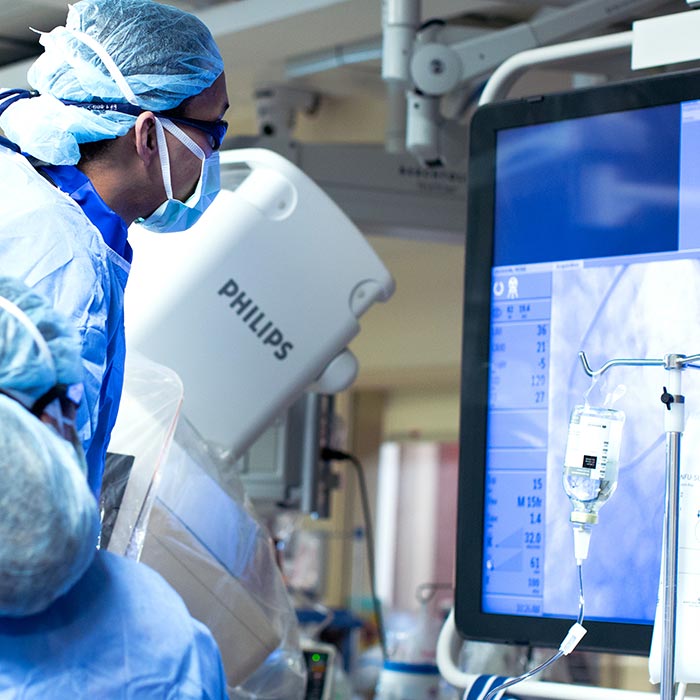
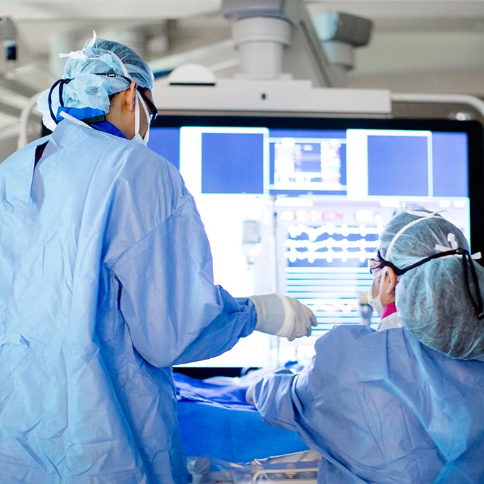
A lot of the input into a TAVR meeting relies on high-quality imaging, and in that respect technology advancements have sped up the march of minimally invasive therapies. Technology has come a long way, and new platforms that bring simplicity to complex procedures have helped interventional radiologists and the physician with their workflows, and created an environment that enables them to accomplish treatment and diagnostic tasks easier. “In the older versions of imaging equipment that we and others have used, there are certain things that have to happen in sequence,” says Dr. Katzen. “One example might be if you’re doing a carotid stent or you’re doing some physiologic study where you have to stop and measure. In the past, the operator had to actually stop the procedure and do whatever measurements that needed to be done while everybody waited.” He says through technologies such as Philips Azurion Image Guided Therapy Systems there is now an incredible amount of information available at the clinician’s fingertips, which prevents the need to step away from a procedure. “Things that you would routinely need to scrub out or go outside the room for, would take time. These various patterns of workflow can actually occur simultaneously and in a seamless way, to allow multiple things, multitasking essentially, to occur during the treatment of a patient,” Dr. Katzen adds. “That improves efficiency and throughput.”
Speeding up clinical decisions
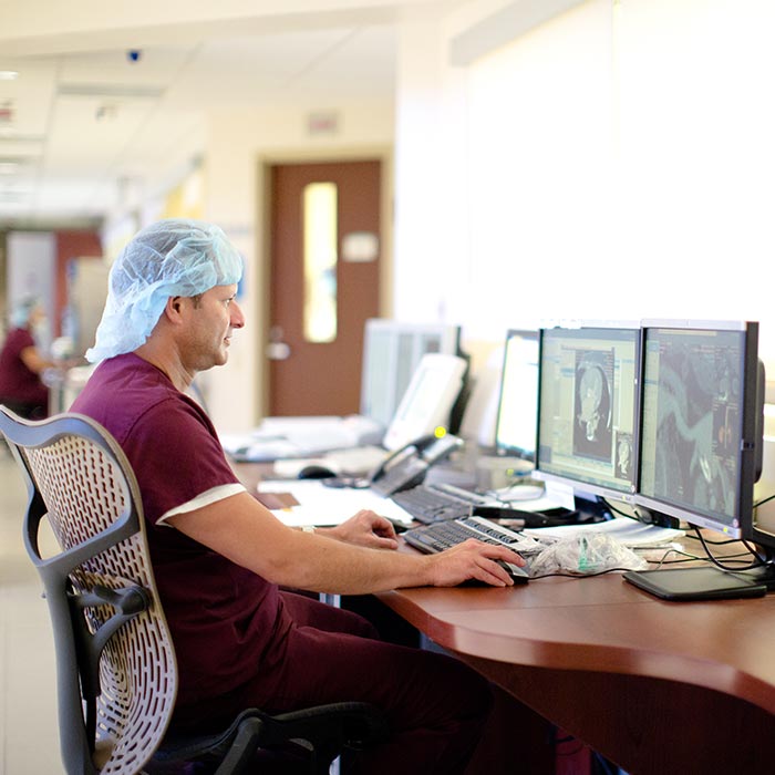
A more seamless flow of information and greater integration of imaging data has led to fewer delays in procedures at the Institute. For interventional radiologists, the ability to bring any type of imaging – whether ultrasound, MR or CT scan – into the Philips IntelliSpace Portal, has helped speed up treatment decisions. “A lot of times when we’re performing procedures, we’re in an arena where we only have one modality. Being able to bring in images from an Ultrasound, CT scan or MRI so that you can use it in real time while you’re performing a procedure is very important as it opens up all of the information while you’re actually performing that procedure,” says Constantino Peña, M.D., an interventional radiologist and Medical Director of Vascular Imaging at the Institute. “The ability to use all of those imaging modalities and integrate them in a platform, in a backbone such as Philips IntelliSpace Portal, allows you to then manipulate and use all these modalities as you see fit to perform a procedure more effectively,” says Dr. Peña.
From hospital to home
Ultimately, it is the patient that benefits most, as their hospital stay can be reduced. A TAVR procedure is not without risks, but it can provide beneficial treatment options to people who may not have been candidates for them a few years ago, while also providing the added bonus of a faster recovery in some cases. Before such procedures came along, length of stay in hospital was measured by weeks, but now they may go home in 24, 48 or 72 hours, says Dr. Katzen. “You can take some clinical examples where a patient with an aneurism, for instance, in their abdomen, used to come in and spend two to four days in the ICU after open surgery, seven days in the hospital, up to 30-60 days recovering, and now those patients can actually go home within 48 hours, really be back to normal within two to three weeks, and have much fewer long-term sequelae than they did in this open repair. These are really dramatic changes. Finally, someone who has a heart attack now goes home in a day.”
Less invasive.
Shorter stays.
A customer story from Miami Cardiac & Vascular Institute
One of the advantages of image-guided, minimally invasive therapies has been we've opened a whole realm of new therapies to patients who were too sick to be treated in the past by any techniques."
Founder and Chief Medical Executive Miami Cardiac & Vascular Institute
Barry T. Katzen, M.D.


The ability to use all of those imaging modalities and integrate them in a platform in a backbone such as Philips IntelliSpace Portal, allows you to then manipulate and use all these modalities as you see fit to perform a procedure more efficiently."
Founder and Chief Medical Executive Miami Cardiac & Vascular Institute
Barry T. Katzen, M.D.
Cardiovascular care with Philips Azurion Image Guided Therapy Platform
No bounds.
Better healthcare.
There's always a way to make life better.
DISCLAIMER: Results are specific to the institution where they were obtained and may not reflect the results achievable at other institutions. FOOTNOTES: 1 American Heart Association, ‘What is TAVR?’ 2 BMJ - https://www.bmj.com/content/354/bmj.i5085.full.print


